Mar. 30, 2023
Why Is Toyota Conducting Brain Research: volume 2Physical well-being related to "UNCONSCIOUS INTELLIGENCE"
The Frontier Research Center of Toyota Motor Corporation (Toyota) is exploring at the possibilities created by integrating brain science and engineering: the aim is to generate innovation for future society. As a part of that process, in 2007, Toyota established an organization for comprehensive collaboration, the RIKEN CBS-TOYOTA Collaboration Center (BTCC), with the RIKEN Center for Brain Science (Wako, Saitama Prefecture [RIKEN CBS]). Since then, they have been conducting joint brain research.*1*2
At BTCC, we talked about the latest research with Dr. Shingo Shimoda, who leads the Intelligent Behavior Control Unit, and Toyota researchers Hitoshi Yamada and Yuhei Yamaguchi in Dr. Shimoda's laboratory.
- What type of research is Toyota advancing in collaboration with Dr. Shimoda?
- Yamaguchi
- To achieve Toyota's mission of "mass-producing happiness," BTCC initiatives promote basic research toward increasing human well-being. The joint research with Dr. Shimoda approaches "physical well-being" with a particular focus on the activities of the nerves and muscles in addition to the brain. As an example, imagine that you are driving a car.
- Yamaguchi
- While driving, you will consciously move your body, for example, to turn the steering wheel and step on the brake in response to your surroundings; you never pay attention to "putting tension on your biceps with this amount" or "moving the calf muscles in this way." You have spontaneously learned unconscious bodily movements based on the various physical exercises experienced since you were a baby.
- Yamada
- Each person has their own unique way of using their body unconsciously, and we believe that appropriately understanding and recognizing these characteristics may lead to more comfortable body movements that align with your intentions, resulting in physical well-being. Dr. Shimoda refers to the unconscious body movements "Unconscious Intelligence" and conducts research to exlore the mechanism of this intelligence and the visualization of these movements. When we look at the muscles, which at first seem to be unrelated to brain science, we find an interesting story.
- What specific type of research is being conducted?
- Dr. Shimoda
- Human muscles consist of slow twitch fibers (slow muscles) which have advantages in endurance and fast twitch fibers (fast muscles) exerting a short-time strong force, and these muscles generate a weak electric current (myoelectricity) when the muscles develop strength. An electromyograph can be used to visualize muscle activity by measuring this weak electric current. We have developed a technology in our research that can distinguish the types of analyzed myoelectricity originating from slow muscles and fast muscles, which allows us to estimate which muscle type primarily provides muscle strength (as shown below).
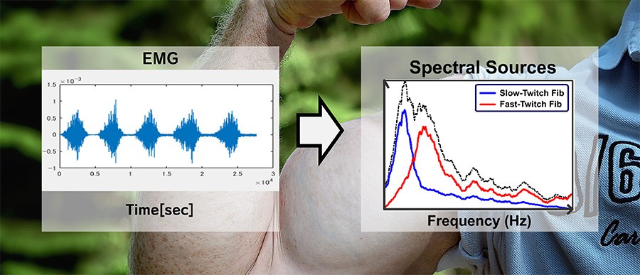
To determine whether myoelectric activity originates from 2 types of muscles, slow muscles and fast muscles, it is possible to analyze muscle synergy in order to measure muscle activity with an EMG (electromyogram). Specifically, the activity signals in the slow muscles and fast muscles can be estimated after calculating the discrepancies between values in a muscle synergy model and those in actual muscle activity followed by data assimilation. This analysis allows us to estimate myoelectric signals coming from slow muscle activity (blue line in the right chart) and fast muscle activity (red line in the right chart). In the right chart, the horizontal axis indicates a frequency, and the vertical axis indicates the power spectrum of each frequency. This chart indicates that fast muscle activity has higher frequencies than that of slow muscle activity.
- What have you found in the research using this technology?
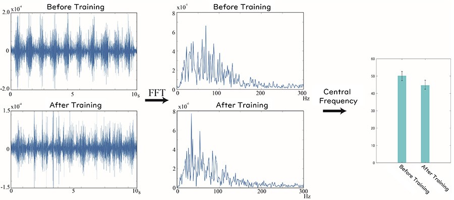
Electromyograms (left 2 charts) and power spectrum intensity (middle 2 charts) before and after training. The center frequency decreases along with training-related proficiency (right chart). This means the myoelectric signal frequency is lower after training, indicating that the slow muscles are superior to the fast muscles in post-training muscle movements.
- Dr. Shimoda
- The above figures show electromyograms and the analysis of their results before and after training for acquiring proficiency in a task. The research revealed that the fast muscles tend to be primarily used to move the body with quick corrections of physical movements while learning a new task, and, after proficiency in the task is attained, the slow muscles primarily function for energy-saving and efficient physical movements.
- Yamada
- So you mean that this research found the way muscles are used unconsciously changed before and after training. This may help us visualize a level of proficiency for physical exercises, leading to a more efficient exercise learning system. I am excited that we have come very close to understanding the relationship between the muscles and the central nervous system, the brain.
- I see. It is good that we can figure out the level of exercise-learning proficiency based on visualization of slow and fast muscle usage. I realize again that we often stay unconscious of many things even about our own body.
- Yamada
- We believe that figuring out such muscle motions will raise the motivation and sense of initiative for individuals to learn, which will contribute to human well-being. In the rehabilitation research advanced by Dr. Shimoda, I often saw patients who became motivated by seeing their own electromyograms. Research has demonstrated increased efficacy of rehabilitation in patients who were given feedback via electromyogram while undergoing rehabilitation.*3
- How will this research evolve in the future?
- Dr. Shimoda
- Ideally, we aim to visualize all muscles to accurately understand how they move unconsciously in humans. The currently available electromyographs, however, primarily detect the motion of the muscles close to the skin, and the muscles far from the skin (deep muscles) are difficult to monitor with these devices. In response to this, we have developed a new electromyograph system that can sense the motions of deep muscles using multiple sensors.*4
- What will be achieved with the new electromyograph system?
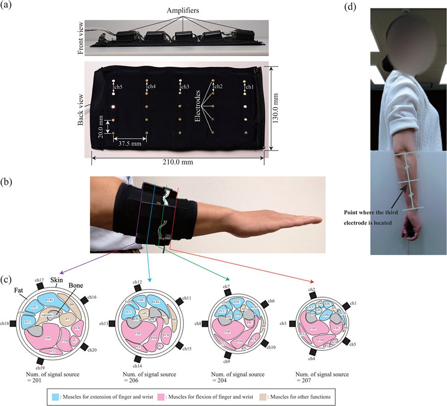
(a) Appearance of the electromyograph system. Amplifiers for signals are implemented on the front side of the system. Active electrodes are placed on the back side (skin-contacting side). (b) Wearing on the arm for use. (c) 5 active electrodes are placed circumferentially around the arm. The blue parts indicate the muscles to extend the fingers and wrist, and the red parts indicate the muscles to flex the fingers and wrist. (d) To keep measurements consistent, the device is worn with the 3rd row of electrodes positioned at the middle point between the elbow and wrist.
- Dr. Shimoda
- This system is used by wrapping it around the arm in the area between the elbow and wrist. Signals are sent from the 5 electrodes (active electrodes) that are placed for electromyographic measurements circumferentially around the arm. The signals are analyzed to determine which muscles generate myoelectricity among the many muscles involved in the arm. The 4 sets of the 5 electrodes are placed in parallel in the area between the elbow and wrist to visualize the muscle motions in the 4 cross sections. The videos shown below indicate the analytic result of the myoelectricities generated when 5 patients open and close there hands with the wrist kept flexed. You can see that the 5 patients seem to make a similar movement, but they use the muscles in different ways. After witnessing these differences, I realized that rehabilitations and various exercises should be customized to seek appropriate methods for each person.
Analytic results for 5 persons are shown in the order from top to bottom. Each row includes 4 cross sections in the area between the wrist and elbow placed in the left to right order. The most left section is close to the wrist, and the most right section is close to the elbow. As you can see, each person shows different usage of the muscles while making similar movements.*4
- The video surely facilitates understanding of the fact that many muscles are used in a complicated way even in a simple movement.
- Yamaguchi
- No other research in the world has developed multipole myoelectric measurements like this using active electrodes that are available for measurements during exercise. We expect this technology to become significant for more effective use of one's own body. In rehabilitation, for example, by becoming conscious of which muscles are not being used compared to healthy physical motions, a patient may undergo more efficient rehabilitation. Additionally, this may provide athletes with more detailed data on which muscles are being used while engaging in their spot. This system may also help professional players determine which muscles should be strengthened and how to move them.
- Dr. Shimoda
- We expect that such research and development will provide fundamental knowledge to correctly understand and more appropriate approach cases where patients or doctors feel things such as "I feel physical things do not work out as intended, today." or "I cannot figure out why this rehabilitation does not work in this patient." Moreover, in the future, we may be able to give predictions such as "Your physical function may change or worsen if you continue your current lifestyle for more several years." Understanding the current and future of one's own body seems to be a key to physical well-being.
- Yamaguchi
- Being conscious of "Unconscious Intelligence" not only contributes to our physical well-being but may also raise our sense of initiative and motivation, finally leading to psychological well-being. In this sense, this research is very attractive.
- Yamada
- This time, we primarily focused on the muscles in the body, and another study is in progress on the question of whether these motions are controlled by the brain or spinal nerves. The spinal cord has seemed to function as a simple reflex; however, we believe that the spinal cord may be involved in a combination of motions. This mechanism itself is considered to be a form of intelligence. This matter was discussed in another volume, which is also worth reading.*1
- We will look forward to future research. Thank you for the very interesting discussion today.
References
| *1 | Gaming in an MRI Machine?! Toyota's Surprise Findings, Toyota Times. |
|---|---|
| *2 | Why Is Toyota Conducting Brain Research: Aiming for Well-Being and Respect for Individuality, Frontier Research Center, Toyota Motor Corporation. |
| *3 | "Self-Support Biofeedback Training for Recovery from Motor Impairment After Stroke", IEEE Access, vol. 8, pp. 72138-72157, 2020, Fady Alnajjar, Ken-ichi Ozaki, Matti Itkonen, Hiroshi Yamasaki, Masanori Tanimoto, Ikue Ueda, Masaki Kamiya, Maxime Tournier, Chikara Nagai, Alvaro Costa Garcia, Kensuke Ohno, Aiko Osawa, Izumi Kondo, and Shingo Shimoda |
| *4 | "Forearm muscle activity estimation based on anatomical structure of muscles", The Anatomical Record, 06 April 2022, Shotaro Okajima, ÁLvaro Costa-García, Sayako Ueda, Ningjia Yang, Shingo Shimoda |
Contact Information (about this article)
- Frontier Research Center
- xr-probot@mail.toyota.co.jp
RELATED CONTENT
MOST POPULAR